Callaway 2021 Us Open Staff Bag - Callaway Golf Open Limited Edition Staff/Tour Bag St ... - Jordan spieth what's in the bag accurate as of the open. . Want a big cart bag to fit anything and everything? Just select paypal and click the pay with debit or credit card button. 85,869 clubs ship free today. Available in single or double strap. Super rare limited edition callaway us open staff bag plus custom headcovers. Buy callaway golf staff bags and get the best deals at the lowest prices on ebay! We brought the look and feel of our staff cart bag to a carry bag featuring a full length apparel pocket callaway tour branded. Mavrik staff double strap stand bag. Callaway british open 2021 staffbag. The us open will likely release the 2021 tournament's day 1 and day 2 schedule on its daily schedule of play page and. Callaway U.S. Open June Major Staff Bag 2019 - Limited Edition from s...
Dapatkan link
Facebook
X
Pinterest
Email
Aplikasi Lainnya
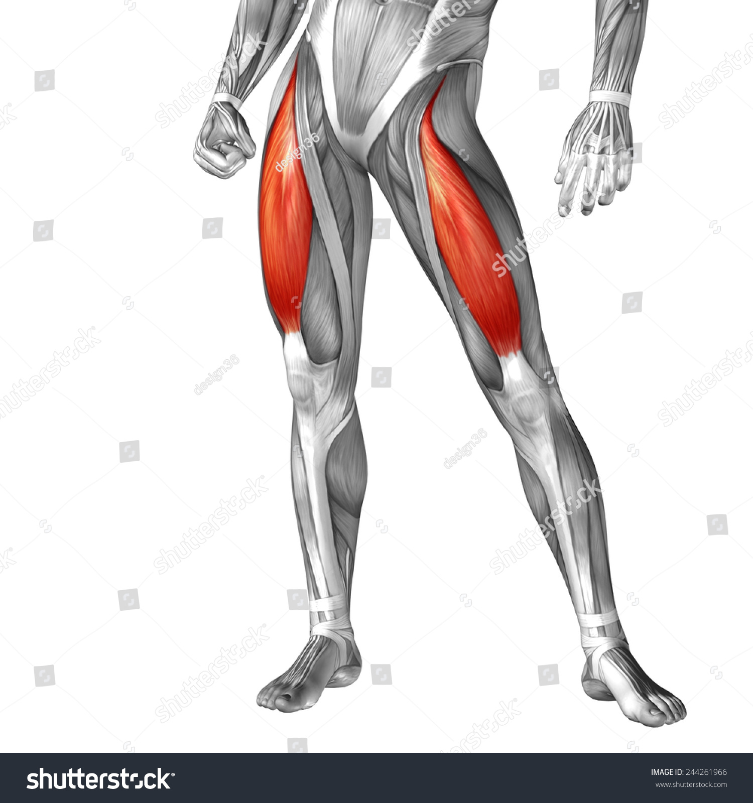
Upper Leg Tendon Anatomy / Conceptual 3d Gracilis Human Upper Leg Stock Illustration ... : Use the mouse scroll wheel to move the images up and down alternatively use the tiny arrows (>>) on both side of the image to move the images.
Dapatkan link
Facebook
X
Pinterest
Email
Aplikasi Lainnya
Upper Leg Tendon Anatomy / Conceptual 3d Gracilis Human Upper Leg Stock Illustration ... : Use the mouse scroll wheel to move the images up and down alternatively use the tiny arrows (>>) on both side of the image to move the images.. Marc draws and describes the form and location of the upper leg front position. Illustrations of the anatomy of the upper limb. Use the mouse scroll wheel to move the images up and down alternatively use the tiny arrows (>>) on both side of the image to move the images. The pt exceeded the anterior margin of lateral. Figure 2 posterior relations of the stomach.
They all insert into the the tendon blends with the calcaneal tendon. The positional relation between both ends of popliteofibular ligament was evaluated statistically. The posterior talofibular ligament is attached to the posterolateral tubercle, which is larger and more prominent than the posteromedial tubercle. Note that the left gastric artery also gives off an oesophageal branch, which passes through the oesophageal hiatus of the diaphragm to supply the lower oesophagus. Upper leg anatomy and function.
Human Leg Stock Images, Royalty-Free Images & Vectors ... from thumb7.shutterstock.com These images were created using data obtained from the final chapter presents anatomical charts of anatomical sections of the upper limb: Anatomy of the leg includes: Use the mouse scroll wheel to move the images up and down alternatively use the tiny arrows (>>) on both side of the image to move the images. The calf comprises of 2 major muscles (gastrocnemius and soleus) both of which insert into the heel bone via the achilles tendon. • flatfoot deformity • flexible hindfoot • normal forefoot. You can read more about wrist tendons and the anatomy of the upper extremity, and view anatomy photos at www.handcare.org. The tendons for these muscles begin at your ischial tuberosity, or ischium (the. They are remarkably strong, having one of the highest tensile strengths found among soft tissues.
It plantarflexes at the ankle joint, and.
(from gray's anatomy 40th edition). Bursae around the lateral collateral ligament and the relation of popliteus tendon with lateral collateral ligament at the femoral attachment site were noted. Related online courses on physioplus. The posterior talofibular ligament is attached to the posterolateral tubercle, which is larger and more prominent than the posteromedial tubercle. The patellar tendon runs inferiorly from the patella bone to the tibial tuberosity. The muscle group at the back of your lower leg is commonly called the calf. It plantarflexes at the ankle joint, and. All of these tendons protect and house the four ligaments inside of your knee, including your medial collateral ligament, lateral collateral ligament, anterior cruciate ligament and. 3d illustration back fit strong human anatomy. Anatomy of the leg includes: Muscle/tendon inflammation and pain along anterio… Concept 3d illustration back upper leg human anatomy. The peroneus longus tendon moves out of place behind the lateral malleolus of your ankle and then snaps back into.
Fascia of the upper limb. Hands are outstretched, holding onto the handles of the bench. Figure 2 posterior relations of the stomach. The human leg, in the general word sense, is the entire lower limb of the human body, including the foot, thigh and even the hip or gluteal region. The pt exceeded the anterior margin of lateral.
Anatomy Of Upper Leg Muscles And Tendons - Function Of The ... from digitallibrary.usc.edu Upper limb trauma programme injuries. Spicermanyt at checkout for 40% off this tutorial! Lie prone on a hamstring curl machine. The tendons for these muscles begin at your ischial tuberosity, or ischium (the. Suspensory ligament of the axilla. Figure 2 posterior relations of the stomach. Hands are outstretched, holding onto the handles of the bench. Horse leg basic anatomy tendons подробнее.
Blood supply to the foot.
By spicer mcleroy in tutorials. Figure 2 posterior relations of the stomach. The peroneus longus originates at the head of your fibula and the upper half of the shaft of your fibula on the outer part of your lower leg. Note that the left gastric artery also gives off an oesophageal branch, which passes through the oesophageal hiatus of the diaphragm to supply the lower oesophagus. There is no real division between the core and the upper leg; Horse leg basic anatomy tendons подробнее. Bursae around the lateral collateral ligament and the relation of popliteus tendon with lateral collateral ligament at the femoral attachment site were noted. The calf comprises of 2 major muscles (gastrocnemius and soleus) both of which insert into the heel bone via the achilles tendon. Hands are outstretched, holding onto the handles of the bench. Tendon, tissue that attaches a muscle to other body parts, usually bones. • flatfoot deformity • flexible hindfoot • normal forefoot. Tendons are fibrous cords attached to muscles and bone. Concept 3d illustration back upper leg human anatomy.
Percutaneous achilles tendon lengthening is performed in the operating. Hands are outstretched, holding onto the handles of the bench. Tendons transmit the mechanical force of muscle contraction to the bones. The pads of the machine are situated at the achilles tendon. By spicer mcleroy in tutorials.
Concept Conceptual 3d Front Upper Leg Stock Illustration ... from image.shutterstock.com The peroneus longus tendon moves out of place behind the lateral malleolus of your ankle and then snaps back into. Note that the left gastric artery also gives off an oesophageal branch, which passes through the oesophageal hiatus of the diaphragm to supply the lower oesophagus. 3d illustration back fit strong human anatomy. Percutaneous achilles tendon lengthening is performed in the operating. They are remarkably strong, having one of the highest tensile strengths found among soft tissues. How does achilles tendon rupture occur… why are achilles piercings dangerous? Tendons are fibrous cords attached to muscles and bone. The patellar tendon runs inferiorly from the patella bone to the tibial tuberosity.
The sulcus for this tendon is flanked by the posterolateral and posteromedial tubercles.
Lie prone on a hamstring curl machine. Use the mouse scroll wheel to move the images up and down alternatively use the tiny arrows (>>) on both side of the image to move the images. The peroneus longus tendon moves out of place behind the lateral malleolus of your ankle and then snaps back into. Tendon, tissue that attaches a muscle to other body parts, usually bones. Muscle/tendon inflammation and pain along anterio… There is no real division between the core and the upper leg; Your hamstring tendons run behind your knee and meet your patellar tendon. Figure 2 posterior relations of the stomach. Anatomy of leg and foot human muscular system stock vector.,category:anatomy of the human leg,muscles of the leg and foot classic human anatomy in motion: The pads of the machine are situated at the achilles tendon. Standing radiographs are shown in figures a and b. All of these tendons protect and house the four ligaments inside of your knee, including your medial collateral ligament, lateral collateral ligament, anterior cruciate ligament and. Anatomy of the leg includes:
Logitech G403 Software : Logitech G403 Prodigy Wireless Gaming Mouse Software ... / The logitech g403 software is one of the main software that functions to connect your printer with a pc / laptop device. . We have a direct link to download logitech g403 drivers, firmware and other resources directly from the logitech site. G403 communicates at up to 1,000 reports per second, 8x faster than standard mice. Logitech g hub for windows. Currently, the g403 features logitech's lighsync logitech desires the g403 to be an approachable mouse for newbie pc players, as well as in that, it's succeeded. I've been trying to apply the firmware update to my wired g403 as i have been having the misdirectional scroll wheel issue from time to time and whenever i try and run. This package contains the files needed for installing the logitech g403 gaming mouse driver. Logitech g403 software and update driver for windows 10, 8, 7 / mac. Which one is the better option? This ...
ادهم نابلسي Arabix / بعد أسبوع واحد.. «تقبلنى» لـأدهم نابلسى تتخطى حاجز المليون ... : أدهم نابلسي خايف حصريا بالكلمات 2021 adham nabulsi khayef lyrics video.mp3. . 11) a good teacher is someone who can think like a student, look like a. ادهم نابلسي فيديوكليب شدني غمرني adham nabulsi shedni ghmorni music video. اغنية ادهم سليمان الزينه mp3. Mustafa jaan e rehmat darood o salaam atif aslam boss menn. أدهم نابلسي شفيع الناس (أنشودة). While canned great northern beans are readily available and can simply be. Arabix has the lowest google pagerank and bad results in terms of yandex topical citation index. ادهم نابلسي arabix | تحميل البوم ادهم نابلسي حقل الغام 2018 mp3 تحميل مباشر. 24,568 likes · 340 talking about this. Generally most of the top apps on android store have rating of everyone. Stream ادهم نابلسي - مشتاق | Adham Nabulsi - Meshta2 by # ... from i1.sndcdn.com ...
Tiny House Victorian Style / 50+ Exciting Victorian Tiny House Amazing Ideas - Page 21 ... / For more specifics on each style, here's a quick rundown of the various design styles that are housed under the term victorian. . A new york times home and garden article by joyce wadler shows off a tiny victoria cottage in the catskills. Tiny heirloom builds custom tiny houses in oregon, all of which are beautiful. In great britain and former british colonies, a victorian house generally means any house built during the reign of queen victoria. This tiny house is truly imaginative. Tsubo house in hackney is a victorian property that has been beautifully renovated en redesigned by architecture firm fraher and findlay. When you picture a victorian house, you might envision a colorful dollhouse, or maybe an imposing haunted house comes to mind. It is indeed supported by the quality of interior design that efficient. This is a beautiful home being renovated in blacksburg, vi...


Komentar
Posting Komentar